What Do Biological Systems Do to Control or Keep the Ph of the Body Stable
Affiliate 11: Introduction to the Body's Systems
11.1 Homeostasis and Osmoregulation
Learning Objectives
Past the end of this section, you will be able to:
- Explicate the concept of homeostasis
- Describe thermoregulation of endothermic and ectothermic animals
- Explain how the kidneys serve as the main osmoregulatory organs in the man torso
Homeostasis refers to the relatively stable land inside the body of an beast. Brute organs and organ systems constantly suit to internal and external changes in guild to maintain this steady land. Examples of internal conditions maintained homeostatically are the level of claret glucose, body temperature, claret calcium level. These conditions remain stable because of physiologic processes that consequence in negative feedback relationships. If the blood glucose or calcium rises, this sends a betoken to organs responsible for lowering blood glucose or calcium. The signals that restore the normal levels are examples of negative feedback. When homeostatic mechanisms fail, the results tin be unfavorable for the animate being. Homeostatic mechanisms keep the torso in dynamic equilibrium by constantly adjusting to the changes that the trunk's systems run across. Even an animal that is plainly inactive is maintaining this homeostatic equilibrium. Two examples of factors that are regulated homeostatically are temperature and water content. The processes that maintain homeostasis of these two factors are chosen thermoregulation and osmoregulation.
Homeostasis
The goal of homeostasis is the maintenance of equilibrium around a specific value of some aspect of the body or its cells chosen a set point. While at that place are normal fluctuations from the set signal, the body's systems volition usually endeavour to go back to this point. A modify in the internal or external surround is chosen a stimulus and is detected past a receptor; the response of the system is to adjust the activities of the system then the value moves back toward the set point. For example, if the trunk becomes too warm, adjustments are made to absurd the animate being. If glucose levels in the blood ascension afterwards a meal, adjustments are made to lower them and to get the nutrient into tissues that need it or to shop it for later use.
When a alter occurs in an animal's environment, an aligning must exist made so that the internal environment of the trunk and cells remains stable. The receptor that senses the alter in the surround is part of a feedback mechanism. The stimulus—temperature, glucose, or calcium levels—is detected by the receptor. The receptor sends data to a control heart, ofttimes the encephalon, which relays appropriate signals to an effector organ that is able to crusade an appropriate alter, either up or down, depending on the information the sensor was sending.
Thermoregulation
Animals can be divided into two groups: those that maintain a abiding body temperature in the confront of differing ecology temperatures, and those that have a body temperature that is the aforementioned as their environment and thus varies with the environmental temperature. Animals that do not take internal control of their body temperature are chosen ectotherms. The trunk temperature of these organisms is by and large like to the temperature of the environs, although the private organisms may practice things that keep their bodies slightly beneath or higher up the environmental temperature. This can include burrowing underground on a hot mean solar day or resting in the sunlight on a cold day. The ectotherms have been chosen cold-blooded, a term that may not apply to an animate being in the desert with a very warm body temperature.
An animal that maintains a abiding body temperature in the face of ecology changes is chosen an endotherm. These animals are able to maintain a level of activity that an ectothermic animal cannot because they generate internal heat that keeps their cellular processes operating optimally fifty-fifty when the environment is cold.
Concept in Action

Watch this Discovery Channel video on thermoregulation to see illustrations of the procedure in a variety of animals.
Animals conserve or dissipate oestrus in a variety of ways. Endothermic animals accept some form of insulation. They have fur, fatty, or feathers. Animals with thick fur or feathers create an insulating layer of air between their skin and internal organs. Polar bears and seals live and swim in a subfreezing surroundings and still maintain a abiding, warm, trunk temperature. The arctic pull a fast one on, for example, uses its fluffy tail every bit extra insulation when information technology curls upwardly to slumber in cold conditions. Mammals can increment body heat production by shivering, which is an involuntary increase in muscle activity. In addition, arrector pili muscles tin can contract causing individual hairs to stand up when the private is cold. This increases the insulating effect of the pilus. Humans retain this reaction, which does non accept the intended effect on our relatively hairless bodies; it causes "goose bumps" instead. Mammals use layers of fat equally insulation also. Loss of meaning amounts of body fat will compromise an individual's power to conserve estrus.
Ectotherms and endotherms utilise their circulatory systems to aid maintain body temperature. Vasodilation, the opening upwardly of arteries to the skin past relaxation of their smooth muscles, brings more blood and heat to the body surface, facilitating radiation and evaporative heat loss, cooling the body. Vasoconstriction, the narrowing of claret vessels to the peel by wrinkle of their polish muscles, reduces blood flow in peripheral blood vessels, forcing claret toward the core and vital organs, conserving heat. Some animals accept adaptions to their circulatory system that enable them to transfer heat from arteries to veins that are flowing next to each other, warming blood returning to the heart. This is called a countercurrent heat exchange; it prevents the cold venous blood from cooling the centre and other internal organs. The countercurrent adaptation is establish in dolphins, sharks, bony fish, bees, and hummingbirds.
Some ectothermic animals use changes in their behavior to help regulate body temperature. They just seek libation areas during the hottest part of the day in the desert to keep from getting as well warm. The same animals may climb onto rocks in the evening to capture estrus on a cold desert night before entering their burrows.
Thermoregulation is coordinated by the nervous organization (Effigy eleven.2). The processes of temperature control are centered in the hypothalamus of the avant-garde animal brain. The hypothalamus maintains the set indicate for body temperature through reflexes that cause vasodilation or vasoconstriction and shivering or sweating. The sympathetic nervous system under control of the hypothalamus directs the responses that effect the changes in temperature loss or proceeds that return the body to the set up point. The set point may be adjusted in some instances. During an infection, compounds called pyrogens are produced and circulate to the hypothalamus resetting the thermostat to a higher value. This allows the torso'due south temperature to increase to a new homeostatic equilibrium point in what is commonly chosen a fever. The increment in torso oestrus makes the torso less optimal for bacterial growth and increases the activities of cells so they are better able to fight the infection.
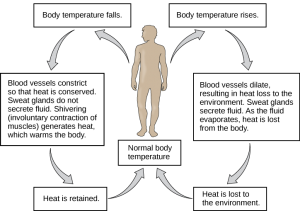
When bacteria are destroyed by leukocytes, pyrogens are released into the blood. Pyrogens reset the body's thermostat to a higher temperature, resulting in fever. How might pyrogens cause the trunk temperature to rise?
<!–Pyrogens increase body temperature past causing the blood vessels to constrict, inducing shivering, and stopping sweat glands from secreting fluid.–>
Osmoregulation
Osmoregulation is the procedure of maintaining salt and water rest (osmotic residual) beyond membranes within the body. The fluids inside and surrounding cells are composed of water, electrolytes, and nonelectrolytes. An electrolyte is a compound that dissociates into ions when dissolved in water. A nonelectrolyte, in contrast, does not dissociate into ions in water. The body's fluids include blood plasma, fluid that exists within cells, and the interstitial fluid that exists in the spaces between cells and tissues of the body. The membranes of the torso (both the membranes around cells and the "membranes" made of cells lining trunk cavities) are semipermeable membranes. Semipermeable membranes are permeable to sure types of solutes and to water, merely typically jail cell membranes are impermeable to solutes.
The body does not exist in isolation. There is a constant input of water and electrolytes into the system. Excess water, electrolytes, and wastes are transported to the kidneys and excreted, helping to maintain osmotic balance. Insufficient fluid intake results in fluid conservation by the kidneys. Biological systems constantly interact and commutation h2o and nutrients with the environment by mode of consumption of nutrient and h2o and through excretion in the form of sweat, urine, and feces. Without a mechanism to regulate osmotic force per unit area, or when a disease amercement this mechanism, there is a tendency to accrue toxic waste and water, which tin can have dire consequences.
Mammalian systems take evolved to regulate non only the overall osmotic pressure across membranes, just too specific concentrations of important electrolytes in the iii major fluid compartments: blood plasma, interstitial fluid, and intracellular fluid. Since osmotic force per unit area is regulated past the move of h2o across membranes, the volume of the fluid compartments can likewise change temporarily. Since blood plasma is 1 of the fluid components, osmotic pressures have a direct begetting on claret force per unit area.
Excretory System
The human excretory system functions to remove waste from the body through the pare equally sweat, the lungs in the form of exhaled carbon dioxide, and through the urinary arrangement in the course of urine. All iii of these systems participate in osmoregulation and waste removal. Here we focus on the urinary system, which is comprised of the paired kidneys, the ureter, urinary bladder and urethra (Figure 11.3). The kidneys are a pair of bean-shaped structures that are located just below the liver in the body crenel. Each of the kidneys contains more than a million tiny units called nephrons that filter blood containing the metabolic wastes from cells. All the blood in the human body is filtered most 60 times a day by the kidneys. The nephrons remove wastes, concentrate them, and form urine that is collected in the float.
Internally, the kidney has iii regions—an outer cortex, a medulla in the middle, and the renal pelvis, which is the expanded cease of the ureter. The renal cortex contains the nephrons—the functional unit of measurement of the kidney. The renal pelvis collects the urine and leads to the ureter on the outside of the kidney. The ureters are urine-begetting tubes that exit the kidney and empty into the urinary bladder.
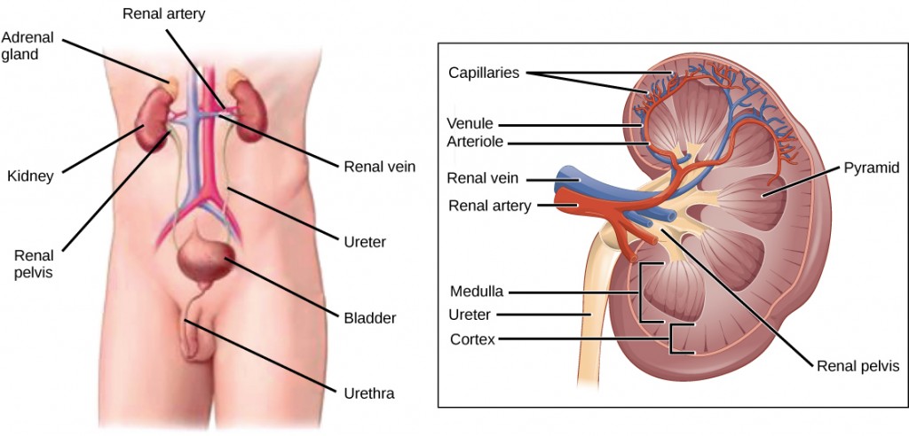
Blood enters each kidney from the aorta, the main avenue supplying the trunk beneath the heart, through a renal artery. It is distributed in smaller vessels until information technology reaches each nephron in capillaries. Within the nephron the blood comes in intimate contact with the waste material-collecting tubules in a structure chosen the glomerulus. H2o and many solutes present in the blood, including ions of sodium, calcium, magnesium, and others; as well as wastes and valuable substances such as amino acids, glucose and vitamins, exit the blood and enter the tubule organisation of the nephron. As materials pass through the tubule much of the h2o, required ions, and useful compounds are reabsorbed dorsum into the capillaries that environment the tubules leaving the wastes behind. Some of this reabsorption requires active ship and consumes ATP. Some wastes, including ions and some drugs remaining in the blood, lengthened out of the capillaries into the interstitial fluid and are taken up by the tubule cells. These wastes are and then actively secreted into the tubules. The claret and so collects in larger and larger vessels and leaves the kidney in the renal vein. The renal vein joins the inferior vena cava, the chief vein that returns blood to the heart from the lower torso. The amounts of water and ions reabsorbed into the circulatory system are carefully regulated and this is an important way the body regulates its water content and ion levels. The waste product is collected in larger tubules and then leaves the kidney in the ureter, which leads to the bladder where urine, the combination of waste material materials and h2o, is stored.
The bladder contains sensory nerves, stretch receptors that betoken when it needs to be emptied. These signals create the urge to urinate, which can be voluntarily suppressed upwardly to a limit. The conscious decision to urinate sets in play signals that open up the sphincters, rings of smoothen muscle that close off the opening, to the urethra that allows urine to flow out of the float and the body.
Dialysis Technician
Dialysis is a medical process of removing wastes and excess water from the blood by diffusion and ultrafiltration. When kidney function fails, dialysis must be done to artificially rid the body of wastes and fluids. This is a vital process to go along patients live. In some cases, the patients undergo artificial dialysis until they are eligible for a kidney transplant. In others who are non candidates for kidney transplants, dialysis is a lifelong necessity.
Dialysis technicians typically work in hospitals and clinics. While some roles in this field include equipment development and maintenance, most dialysis technicians work in direct patient care. Their on-the-job duties, which typically occur under the direct supervision of a registered nurse, focus on providing dialysis treatments. This can include reviewing patient history and electric current condition, assessing and responding to patient needs before and during treatment, and monitoring the dialysis process. Handling may include taking and reporting a patient's vital signs, preparing solutions and equipment to ensure accurate and sterile procedures.
Section Summary
Homeostasis is a dynamic equilibrium that is maintained in trunk tissues and organs. It is dynamic considering information technology is constantly adjusting to the changes that the systems encounter. It is an equilibrium considering body functions are kept within a normal range, with some fluctuations around a set indicate. The kidneys are the master osmoregulatory organs in mammalian systems; they part to filter blood and maintain the dissolved ion concentrations of body fluids. They are made up internally of three distinct regions—the cortex, medulla, and pelvis. The blood vessels that ship blood into and out of the kidneys ascend from and merge with the aorta and inferior vena cava, respectively. The nephron is the functional unit of the kidney, which actively filters blood and generates urine. The urine leaves the kidney through the ureter and is stored in the urinary bladder. Urine is voided from the body through the urethra.
Glossary
ectotherm: an organism that relies primarily on environmental heat sources to maintain its body temperature
endotherm: an organism that relies primarily on internal estrus sources to maintain its torso temperature
interstitial fluid: the fluid found betwixt cells in the body, similar in constitution to the fluid component of blood, but without the loftier concentrations of proteins
kidney: the organ that performs excretory and osmoregulatory functions
nephron: the functional unit of measurement of the kidney
osmoregulation: the mechanism by which h2o and solute concentrations are maintained at desired levels
osmotic balance: the appropriate values of water and solute concentrations for a healthy organism
renal artery: the avenue that delivers claret to the kidney
renal vein: the vein that drains blood from the kidney
fix bespeak: the target value of a physiological land in homeostasis
ureter: the urine-bearing tubes coming out of the kidney
urethra: the tube that conducts urine from the urinary float to the external environment
urinary float: the structure that the ureters empty the urine into the advisable values of water and solute concentrations for a healthy organism
Source: https://opentextbc.ca/biology/chapter/11-1-homeostasis-and-osmoregulation/
0 Response to "What Do Biological Systems Do to Control or Keep the Ph of the Body Stable"
Post a Comment